
The breast is an exocrine gland that develops during the life of women. It contains the mammary glands, which become active during periods of lactation to produce milk. In addition to breast-feeding, the breast plays an important role in the shape and confidence of a woman. However, the breast is subject to many diseases, among which,is cancer.
Breast cancer is a malignant tumor that develops in the mammary gland, especially in the inner lining of the milk ducts. This condition occurs when abnormal cells, under the action of an external aggression or carcinogens (such as smoking or ultraviolet rays), multiply anarchically to form a malignant growth. Without a curative intervention, these malignant cells will continue to proliferate, and spread into other tissues in the body to form metastatic cancers. This condition is called metastatic breast cancer. Symptoms or characteristic of metastatic breast cancer depend greatly on the location of the secondary tumor.
Although men are also victims of breast cancer, the disease is more common among women. It is estimated that about 1 woman out of 7 will be affected by breast cancer during their lifetime, which makes the disease the leading cause of death among gynecological cancers in developed countries.
Breast Cancer Statistics
Breast cancer is the most common form of cancer in women. In spite of medical advances, it remains a deadly disease all over the world, killing about 519,000 people worldwide in 2004. In the US alone, breast cancer kills nearly 41,116 women and 375 men each year. According to the National Cancer Institute (NCI), it is estimated that 192,370 women were diagnosed with breast cancer in 2009 with about 40,170 women dying of it.

However, the incidence of breast cancer varies with age and race; from 2002-2006, the median age at diagnosis for breast cancer was 61 years of age. Age and percentage of women diagnosed with breast cancer during 2002-2006 were approximately:
- 0.0% for women under age 20
- 1.9% for women between 20-34
- 10.5% for women between 35-44
- 22.5% for women between 45-54
- 23.7% for women between 55-64
- 19.6% for women between 65-74
- 16.2% for women between 75-84
- 5.5% for women aged 85 years or older
Incidence and death rates by race
| Incidence | Death | |
| White | 127.8 per 100,000 women | 23.9 per 100,000 women |
| Black | 117.7 per 100,000 women | 33.0 per 100,000 women |
| Asian/Pacific Islander | 89.5 per 100,000 women | 12.5 per 100,000 women |
| American Indian/Alaska Native | 74.4 per 100,000 women | 17.6 per 100,000 women |
| Hispanic | 88.3 per 100,000 women | 15.5 per 100,000 women |
| All Races | 123.8 per 100,000 women | 24.5 per 100,000 women |
Breast Cancer Causes
The breast is an organ composed mainly of fat, glands and milk ducts (also called lactiferous ducts, galactophorous ducts, mammary ducts, or mammillary ducts). During periods of lactation, the glands produce milk, which is transported to the nipple by the ducts. In a healthy breast, cells group together to form tissues. Each tissue works togetherto perform a similar function. The breast tissue is influenced by hormones like estrogen and progesterone. These two hormones are produced by women in variable amounts throughout puberty, pregnancy, and lactation.
Breast cancer occurs when a group of normal cells begin to transform and divide in an uncontrolled manner to become malignant. If your immune system is healthy enough, these tumor cells will be destroyed. Otherwise, these diseased cells continue to multiply to form a malignant tumor (cancer) within the breast. These cancer cells can remain in the breast asymptomatically for months or years. With time, they travel through the lymphatic system or bloodstream to invade other organs distant from the breast to form new tumors called metastases.
Despite advances in medical science, the exact causes of breast cancer are still not well known. However, many conditions are suspected in the development of the disease. For example, immediate family history of breast cancer represents a 5-10% risk factor of the disease. In addition, excess weight, diet rich in animal fats, lack of pregnancy, late first pregnancy, early onset of menstruation, late menopause, certain forms of mastopathy (any non malignant disease or pain of the mammary gland), and malignant tumor of the large bowel, uterine, or ovaries also seem to contribute in the development of breast cancer.
Breast Cancer Risk Factors
About 70% of breast cancer cases occur without any explicit cause. However, there are factors clearly identified as obvious risks of breast cancer. Most factors that can increase the risk of breast cancer include:
 Age: Although breast cancer can affect young women of all ages, its risk increases with age. It is shown that breast cancer is more common among older people. Therefore, if you are aged 45 or older, your risk of developing breast cancer may be about 2 times higher than those in their thirties; if you are between 55 and 64, your risk is about 3 times higher than women in their forties.
Hormone replacement therapy (HRT): According to WHO (World Health Organization), menopause hormone therapy (MHT) is a risk factor for breast cancer especially when taken for more than five years. some scientists believe that the risk of developing breast cancer increases by 1% per year by taking estrogen alone and 8% per year if the therapy consists of a combination of estrogen and progesterone. In addition, studies show that hormone therapy can also increase the risk of colon cancer. However, those risks may disappear about two years after cessation of the therapy.
Prolonged exposure to endogenous estrogen: In menopausal women, this hormone is produced by the body under the action of the adrenal glands. In premenopausal women, it is produced at 60% by the ovaries (estradiol) and 40% by the adrenal glands (estrone). Prolonged exposure to endogenous estrogens may be a risk of breast cancer. It is found a higher rate of breast cancer among women 35 to 65 years old who had their first menstruation early (before age 12 for instance), went into late menopause, nulliparity (no pregnancy), or experienced late pregnancy.
Family history: Your genetic factors to develop breast cancer are about 5-10% if you have close relatives suffering from the disease. Genetics is primarily responsible for breast cancers occurring before age forty. Some genes that appear responsible for developing the disease include BRCA I—a defect on chromosome 17 associated with an increased risk for breast cancer, and inherited by only 1 in 200 women—and BRCA II which is adefect on chromosome 13 which is associated with an increased risk of ovarian cancer, fallopian tube cancer, prostate cancer, and pancreatic cancer, as well as malignant melanoma. BRCA II is also associated with breast cancer in men.
It is also thought that defects in TSG101 (tumor susceptibility gene 101) may have a role in the development of a breast tumor. However, the significance of TSG101 alterations in development of cancer (carcinogenesis) is controversial since aberrant transcripts of TSG101 gene have also been identified in normal non-cancerous tissues.
Ataxia-telangiectasia , a rare neurodegenerative disorder, is another inherited disease suspected to weaken the immune system, leading to respiratory disorders and increased risk of breast cancer.
Nutrition : In addition to its beneficial effect on bone and against chronic pain, vitamin D may play a role in the prevention of the proliferation of cancer cells in women’s breast while Vitamin A deficiency among female victims of breast cancer seems to contribute to the development of the disease. A group of doctors at Mount Sinai Hospital in Toronto, compared the vitamin D levels of 760 women with breast cancer with those of 1,140 healthy women. The studies showed that high levels of vitamin D were associated with a 24% decreased risk of breast cancer.
It has been discovered that soy products, some fatty acids (mostly omega-3), fruits, cruciferous vegetables, and other natural foods that are rich in anti-oxidant vitamins can reduce up to 20% the risk of breast cancer. These substances fight against breast cancer by destroying free radicals and blocking the hormone receptors.
Smoking: In addition to pulmonary, oral, head and neck cancers, cigarette smoke can also cause breast cancer. Comparably to non-smoker women, it is shown in many studies that the risk of breast cancer before age fifty is about 70% higher among women who start smoking regularly within five years after the onset of their menstruation.
Alcohol: Even moderate consumption of alcohol is a causative factor for breast cancer. This risk increases by 9% for each glass of alcohol consumed daily. Contribution of alcohol in the development of breast cancer is foundin pre-menopausal women and in postmenopausal women who take hormone replacement.
X-rays and mammography: The modern mammography equipment delivers very little radiation compared to what existed 15-20 years ago, which lowers considerably its carcinogenic potentiality. However, the risk of breast cancer exists for women less than thirty years of age due to glandular susceptibility and the fact women at these ages require greater amount of radiation for imaging of their breasts. Therefore, repeated mammography can increase the risk of breast cancer in young women.
Puberty and menopause – Early puberty and late menopause can increase the susceptibility for breast cancer. A woman whose menopause happens naturally after age fifty-five has more chances to have breast cancer than a woman who has her menopause before age forty-five.
Pregnancy – The absence of pregnancy can increase the chances of breast cancer. In addition, if you have your first pregnancy in your thirties, you are two times more likely to develop breast cancer over a woman who becomes a mother in her early twenties. The risk is even higher if you have no children.
Breastfeeding – Breastfeeding plays a key role in preventing breast cancer. Studies have shown that prolonged breastfeeding reduces the risk of developing breast cancer. In addition, it provides many benefits in the physical and mental development of the infant. By breastfeeding you increase the chance of your infant to be healthy, and decrease your risk of developing breast cancer.
Breast Cancer Symptoms & Signs
At the early stage of the disease, the majority of women with breast cancer have no signs or symptoms that impact their health. Some women may feel “healthy” for months or even years while the tumor is already in their breast. In advanced stages, symptoms emerge. Symptoms of breast cancer are highly variable and depend mainly on the location and extent of the tumor. In general, breast cancer symptoms may include:
- weight loss
- loss of appetite
- redness or retraction of the skin of the breast
- yellowing of the “white of the eye (icterus)
- change in the size or shape of the breast
- inflammation and increased warmth in the breast
- back pain, which may indicate bone metastasis
- peeling or flaking of the nipple skin – sometimes accompanied by bloody discharge
- irritating cough accompanied by shortness of breath – which may indicate lung metastases
Note: If you experience these symptoms, it does not mean you have breast cancer. These symptoms can be caused by medical conditions other than cancer. Only your doctor can confirm your breast cancer diagnosis.
Breast Cancer Diagnosis
There are several methods of screening for breast cancer: breast self-examination (BSE), magnetic resonance imaging (MRI), ultrasound, estrogen, progesterone receptor tests, genetic testing, mammography every 1-2 years, and biopsy.
Breast self-examination (BSE) – A monthly breast self-exam is the easiest way to find breast cancer at an early stage. Women who practice regular self-breast examination have more chances to discover a smaller and less developed breast cancer than those who do not. They have more chance of being cured or live longer with the disease. You can start practicing regular self-exam from the age of twenty. By doing so, you will become familiar with specific texture of your own breasts and know how to discover precancerous abnormalities or early cancerous growth at an earlier stage.
It is recommended to do a breast self-exam during non hormonal stimulation, or 7 to 10 days after the end of your menstruation. If you are irregular or postmenopausal, you can chose a specific date to do the test. During the examination, look for the following things:
- nipple discharge
- sensation of a lump or thickening of the breast
- sensation of a nodule in the armpit or neck
- localized retraction of the skin (like an orange peel) or the nipple
- persistent and painful inflammation of the skin of the breast
- eczema, redness or other abnormalities in the nipple
- visible or palpable change of breast
Although you should always take seriously any recent change in appearance of your mammary gland, a palpable nodule is not necessarily cancer. It may be another breast disease such as mastitis, cyst or fibroadenoma, which is the most common noncancerous breast tumor in young women. A change in your breast may also be due to hormones. Consult your doctor.
Clinical breast exam – Your “self-breast exam” is not always enough to detect abnormalities in your breast; you may need a breast examination by your doctor. The American Cancer Society recommends this test once every three years until the age of forty for even women who are not at risk. The monthly self breast exam, clinical breast examination (by your doctor), and mammography are three key tests in early detection of breast cancer.
Mammogram – This medical technique is often performed in breast cancer diagnosis. It allows your physician to study your mammary gland and possibly detect anomalies, lesions, and breast cancer even at an early stage. This examination is recommended when the breasts are not congested due to menstrual cycle as the breasts are less sensitive to pressure from the mammogram and require lower doses of x-rays. The mammogram test is the most reliable in detecting breast cancer at a very early stage. In fact, early mammography can increase your chance of healing up to 90%.
Other tests such as clinical palpation, ultrasound, scintigraphy, CT scan, and magnetic resonance are often complementary to mammography and can in no way replace it. Although the risk of developing breast cancer due to mammogram is negligible, excessive use can contribute to the development of breast cancer.
Digital mammography (Senographe 2000D) – This screening method is a recent development, but effective. Invented by General Electric, Senographe 2000D was approved by the FDA in January 2000 for the diagnosis of breast cancer. Digital mammography can detect breast cancers even when they are not palpable, but already manifested by fine calcifications or discrete changes in the anatomy of the breast. The image obtained with the Senographe 2000D is often better than standard imaging techniques.
Computer-aided detection (CAD) – This radiological procedure is a recent advance in the diagnosis of breast cancer. It highlights micro calcification clusters and hyperdense structures in the soft tissue of the breast. However, CAD seems less effective in detecting early breast cancer than the digital mammography; therefore, it is often used along with a mammogram. In addition, the CAD is more sensitive for microcalcifications than for masses.
Scintigraphy – Scintigraphy is a medical technique that uses radioactive materials to produce images of the breast. This examination is very specific in detecting malignancy, but it cannot detect lesions that are not larger than 1 cm. Scintigraphy presents no risk to your health because the injected fluid is quickly excreted in your urine. Scintigraphy is complementary to mammogram and is recommended:
1) when the mammogram indicates the possibility of a malignancy
2) in cases of dense breasts that make difficult the interpretation of the mammogram
3) when the results of the needle biopsy are not sufficiently conclusive – showing little cancerous cells
4) in case the patient has had cosmetic surgery or breast implants
5) in case of complications of a local cancer following a chemotherapy
6) if during the palpitation, your doctor has discovered a mass that is not visible on mammography
Magnetic resonance imaging (MRI) – Emerging in the early 1980s, MRI have become an important tool in the diagnosis of many diseases including breast cancer. It is based on the principle of nuclear magnetic resonance (NMR), which allows your doctor to take pictures of the soft tissue of your breast. Unlike a CT-scan, the MRI is a non-invasive procedure that does not radiate. It can visualize small sized-cancers through their neovascularization.

MRIs are widely used in the diagnosis of tumors and play an active role in many forms of biopsy. A MRI helps your doctor monitor the evolution of the cancer during and after treatment. However, MRIs can give false-positive results; therefore, it is often used in addition to mammography.
Breast ultrasound – A breast ultrasound is a medical procedure that allows your doctor to obtain images of your breasts by using sound waves of high frequency. These images give your doctor the possibility to evaluate abnormalities found during the mammogram or clinical exam. A breast ultrasound involves no radiation and poses no health problem.
Biopsy – The imaging techniques can reveal a cancerous mass, but they can’t confirm the presence of cancer cells in your breast. Your physician will perform a biopsy to accurately confirm the diagnosis. In general, the biopsy consists of removing a sample from the breast tissue or cells where the tumor is suspected. The sample will be microscopically studied to obtain accurate information on the overall structure of the fragment removed. Biopsy is often associated with a bacteriological, immunological or biochemical study. There are different types of biopsy that your doctor can perform:
- Thin Needle aspiration biopsy(NAB) – Also called fine needle aspiration cytology, NAB is a simple and fast procedure, yet reliable in detecting cancer cells. Usually, the pathologist uses a thin needle to extract tissue or fluid samples from the lump in the breast to examine under microscope. The entire procedure can last 30 minutes or less.
- Large needle aspiration biopsy (LNAB) – This procedure is nearly the same as the fine needle aspiration cytology. The difference is that the needle is bigger and it takes more tissue for analysis, which allows a definitive diagnosis.
- Surgical biopsy – Performed under local or general anesthesia, a surgical biopsy is the most reliable method to accurately confirm a breast cancer diagnosis. The surgeon removes either a portion of the tumor (incisional biopsy) or the entire tumor plus the surrounding tissue (excisional biopsy) to allow a pathologist to do a microscopic examination. After analysis, your oncologist will be able to determine if your condition is cancer or a benign tumor.
- Stereotactic biopsy – This is a painless removal of samples from a lump in your breast visible only by mammogram or ultrasound. During the procedure, your pathologist will use a special computer to guide the needle to the lump from which he will take the samples. It is an outpatient procedure done under local anesthesia, which can replace surgical biopsy with comparable reliability. A stereotactic biopsy can be performed by a certified technologist, but it requires interpretation of a physician or a board certified radiologist.
Breast Cancer Stages
Now that the cancer is found in your breast, it is important for your doctor to determine if it has spread to other tissues or organs of your body such as the lymph nodes in the armpit.
Staging tests – Your doctor can use the same exams performed in the diagnosis to determine the stage of the cancer. In some cases, additional tests are needed. Your doctor may remove several lymph nodes for microscopic analysis. After removal, the sample will be sent to a pathologist for microscopic examination to look for signs indicating metastases. If the test is positive, you are diagnosed with metastatic breast cancer.
Knowing the stage of your breast cancer is very important for your oncologist to determine the type of treatment you must undergo. Your doctor will use the stage of the cancer to recommend the treatment that is most appropriate and capable to combat the disease. In general, breast cancer stages include the following:
Breast cancer in situ – This stage of breast cancer is commonly known as stage 0, and it represents 20% or less of breast cancers. There are 2 types of breast carcinoma in situ.
- Lobular carcinoma in situ(LCIS) – LCIS is often detected during a mammogram. At this stage, the tumor is not considered cancerous; the scientists call it carcinoma in situ or stage 0 breast cancer. However, it is taken into consideration, for those who have lobular carcinoma have up to 25% chance of developing breast cancer in the next twenty-five years.
- Ductal carcinoma in situ (DCIS) – Also called intraductal carcinoma, DCIS is a particular form of breast cancer in the very early stages that has not spread beyond the lobule. During this stage, there is no infiltration of cancer cells through the fatty breast tissue or the membranes surrounding the lactiferous. Intraductal carcinoma accounts for nearly 25% of all breast cancer diagnosis. As lobular carcinoma in situ, ductal carcinoma in situ is often detected during a mammogram
Stage I – In this early stage, the size of the tumor is usually 2 centimeters or less and the cancer has not spread outside the breast. If you are diagnosed with a stage 1 breast cancer, you are more likely to be cured.
Stage II – A stage 2 breast cancer presents three different characteristics:
- Stage IIA breast cancer – the size of the tumor remains less than or equal to 2 centimeters but has spread to axillary lymph nodes in the armpits
- Stage IIB breast cancer– the cancer has a size between 2 and 5 cm with or without having spread to lymph nodes in the armpit
- Stage IIC breast cancer – the size of the cancer is more than 5 cm but it has not spread to lymph nodes in the armpit
Stage III – The specialists in ecology divide stage III breast cancer in three substages (categories): stage IIIA, IIIB and IIIC.
- Stage IIIA breast cancer – During stage IIIA, 1) The extension of the cancer is less than 5 centimeters and has spread to axillary lymph nodes and the lymph nodes are connected with each other or with other structures. 2) The size of the cancer is greater than 5 centimeters and has spread to axillary lymph nodes.
- Stage IIIB breast cancer – During stage IIIB, 1) the cancer has spread to nearby tissues of the breast (chest wall, ribs, chest muscles, etc..). 2) The cancer has spread to lymph nodes inside the chest wall. At this stage, your survival chances decrease.
- Stage IIIC breast cancer – The cancer can be of any size, and has spread to: 1) 10 or more lymph nodes under the arm; 2) lymph nodes above or beneath the collarbone and near the neck; 3) lymph nodes under the arm.
Stage IV: This final stage indicates a very advanced breast cancer. The cancer has metastasized to other organs of the body such as bones, lungs, brain, etc. The tumor may be extended locally to the skin and internal lymph nodes in the neck.
Recurrence – A breast cancer is considered recurrent when it returns after treatment. The cancer can develop in the original location or in other sites. In general, a recurrent breast cancer is more difficult to be eradicated; survival chances decrease considerably.
Breast Cancer Treatment
Breast cancer treatment varies depending on the stage or the severity of the tumor at diagnosis. In fact, not only the treatment, even the prognosis depends somehow on the BCSAD (breast cancer stage at diagnosis). Five types of treatment are used to fight breast cancer: chemotherapy, radiotherapy, hormone therapy, biological therapy, and surgery.
Chemotherapy
Chemotherapy is a systemic cancer treatment consisting of strong drugs used to shrink or completely eliminate the tumor in your breast. The drugs travel through your body via the bloodstream to destroy cancer cells. For some breast cancers, chemotherapy is very effective and can completely cure the disease. However, in addition to cancer cells, normal cells are also damaged the chemotherapy, which lead to side effects:
- hair loss
- weight loss
- decreased appetite
- darkened and thickened skin
- blistering skin or acne
- mouth blistering
- dry mouth
- fatigue
- painful urination or red urine
- black, tarry stools
- unusual bruising or bleeding
- cough
- congestion
- fever
- dizziness
- chills
- shortness of breath
- sore throat
- swelling of the feet or ankles
- nausea and vomiting
- skin rash
Chemotherapy drugs can be taken by mouth, intravenously or intramuscularly. At first, chemotherapy drugs approved for the adjuvant treatment of breast cancer were a combination of cyclophosphamide (Cytoxan), methotrexate (Trexall, Rheumatrex), and 5-flurouracil (5-FU). In recent years, however, other drugs were added in the treatment of breast cancer. Among which, there are Adriamycin (doxorubicin) and Taxol (paclitaxel); they seem to bring good results in the fight against breast cancer.
Radiation therapy (Radiotherapy)
Radiation therapy is a medical procedure involving the use of x-rays at high energy to kill cancer cells and shrink the tumor. Unlike chemotherapy, which is systematic, radiotherapy delivers a precise dose of radiation to the volume of the tumor, thus, sparing the maximum surrounding healthy tissue. Choosing the exact dose (amount of energy that radiation will deposit in the tissues) of radiotherapy is very important. According to many recent studies, under dosing beyond 5% is less effective and increases the risk of recurrence. Therefore, your oncologist will recommend the dose most capable of fighting the cancer.
Usually you are administered the therapy 4-5 days per week for a period of 5-6 weeks consecutively. A session of radiation can last 20 minutes or less. The therapy is painless (does not cause pain), but causes side effects, which may include:
- fatigue
- discoloration of the breast
- red lips
- constipation
- vomiting and nausea.
A healthy, well-balanced diet along with medicines can remedy those effects.
During and after the therapies (chemotherapy and radiotherapy), medical surveillance is very important. Some complications can occur with the heart, lung, and digestive system. With the invention of sophisticated and computerized machines, radiotherapy has become less dangerous, and causes fewer side effects. If your doctor thinks it will be helpful, he will recommend an internal radiotherapy (brachytherapy) along with the external radiation therapy
Brachytherapy
Also called internal radiotherapy, sealed source radiotherapy or curietherapy, brachytherapy is based on the same principle as radiotherapy. The difference is that radioactive sources are introduced directly into the cancer, making this method less destructive to healthy surrounding cells of the treated area. In addition, brachytherapy can only be performed to treat non-metastatic cancers. Two main types of brachytherapy can be used in the treatment of breast cancer:
Interstitial: During an interstitial brachytherapy, your oncologist will place the radioactive source directly into your breast. The radioactive material can be in the form of plastic tubes, hollow metal needles, seeds, or wires. These materials are placed under the skin of your breast after receiving local, epidural or general anesthesia.
 Endocavitary: This form of radiotherapy consists of placing the radioactive source in natural cavities of the breast affected by the cancer. Endocavitary irradiation therapy allows your doctor to irradiate the tumor while protecting surrounding organs.
Note: Endocavitary irradiation therapy is in experimental phase; it is not yet approved by the FDA.
Hormone therapy
In some types of cancers such as breast cancer and prostate cancer, the cancer cells depend on hormones to multiply; these types of cancers are called hormone-dependent cancers. Therefore, to stop the proliferation of the cancerous cells, your doctor may recommend you take drugs capable of blocking hormonal stimulation. Along with chemotherapy or radiotherapy, hormone therapy (HT) is commonly used to fight breast cancer. While results vary from one patient to another, hormone therapy is less toxic and causes fewer side effects than chemotherapy.
However, this therapy cannot be used in all types of breast cancer. Some young and pre-menopause women sometimes have cancers that are not hormone receptors and the cancer cells are not sensitive to hormone therapy.
In the treatment of breast cancer, hormone therapy may include two groups of drugs: selective estrogen receptor modulators (SERMs) and aromatase inhibitors.
Selective estrogen receptor modulators (SERMs): This class of drugs acts by blocking the action of estrogen in breast tissue and preventing breast cancer cells to multiply. These drugs act like estrogen on some cells (by stimulating the estrogen receptors), while blocking the effects of estrogen on other cells (by inhibiting the estrogen receptor). SERM has a preventive and curative effect against breast cancer; it kills cancer cells and reverses the growth of the tumor.
Tamoxifen is one of the SERM, which has revolutionized the hormonal treatment of breast cancer. It can be used in the prevention and treatment of breast cancer. Raloxifene is another SERM drug group and has characteristics similar to tamoxifen. However, it has a half-life much shorter than tamoxifen and should be used in higher dose.
Side effects of elective estrogen receptor modulators may include:
- night sweats
- water retention
- weight loss
- irregular menstrual periods
- hot flashes
- vaginal itching, discharge or dryness
Older women may experience serious complications such as:
- bone pain
- back pain
- headaches
- cough
- high cholesterol
- blood clots
- endometrial cancer
Aromatase inhibitors: Aromatase inhibitors are a recent class of drugs used in hormone therapy to treat breast cancer in menopausal women. They act by reducing estrogen levels in the blood of postmenopausal women. They inhibit or inactivate aromatase, an enzyme responsible for the synthesis of estrogens from androgens of adrenal origin. Unlike tamoxifen, users of aromatase inhibitors may experience and increased risk of osteoporosis; they generally are well tolerated by most women. The most common aromatase inhibitors are anastrozole (Arimidex), letrozole (Femara), and exemestane (Aromasin).
Surgical treatment
Although surgery can lead to pain and other adverse effects, if you have breast cancer, your chance of undergoing surgery to remove the cancer is very high. The type of surgery performed depends greatly on the size of the tumor. Your surgeon may also perform an axillary lymph node dissection on the same side as the tumor to determine if the cancer has spread to the lymph nodes. In general, the surgeon will perform one of the following surgical procedures: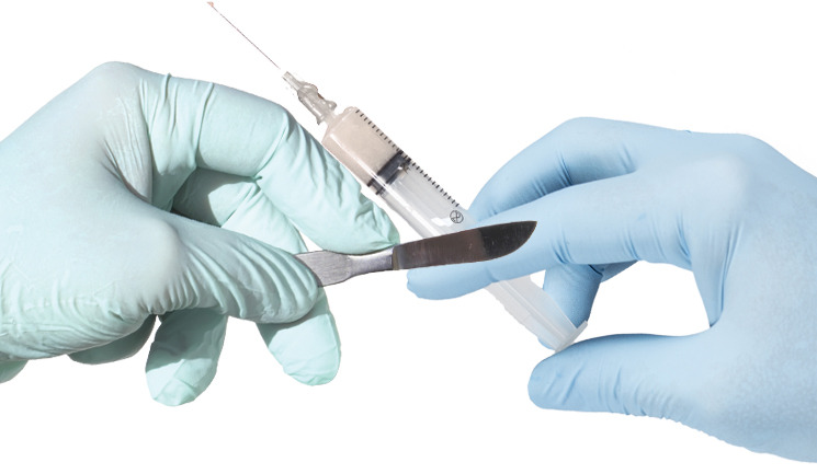
- Lumpectomy: Often performed in surgical treatment of breast cancer, lumpectomy is a surgical procedure consists of removing breast tumor (benign or cancerous) and the tissue that surrounds it. It is usually followed by radiotherapy to kill cancerous cells remaining in the breast tissue. Your surgeon may also remove some axillary lymph nodes during the lumpectomy. However, there are tumors that cannot be eradicated by lumpectomy:
1) large tumor
2) cancer deep within the breast
3) cancer metastasis (in the same breast)
4) Inflammatory breast cancer
If you have already had radiation therapy or suffer from a connective tissue disease, lumpectomy may not be appropriate for you.
Partial mastectomy – Also called segmental mastectomy, partial mastectomy involves surgical removal of the tumor along with a large amount of breast tissue and some skin. Depending on the extension of the tumor, your surgeon may also remove a small part of your chest and some lymph nodes. Partial mastectomy is often followed by radiation therapy.
Simple mastectomy – This surgery is more radical than both lumpectomy and partial mastectomy; it involves complete removal of the breast including the nipple, areola, lobules, ducts, fatty tissue, and skin. A mastectomy may be followed by radiation therapy, chemotherapy, or hormone therapy. After surgery, the surgeon may recommend a breast prosthesis for a date when it is possible to consider a reconstruction.
Modified radical mastectomy: This method is the most performed surgical intervention in the treatment of breast cancer. It involves the removal of the entire breast including the nipple and the skin tissue but without removing the pectoral muscles, which includes the pectoralis major muscle and pectoralis minor muscle. Your surgeon may remove some axillary lymph nodes to determine if the cancer has metastasized.
Radical mastectomy: This is a total removal of the breast including underlying pectoral muscles and axillary lymph nodes. Your surgeon will perform this operation if the cancer is deep within your breast or if the cancer cells have invaded your chest wall.
Reconstructive surgery – If you have a mastectomy, it can negatively change your appearance, and causes emotional and social impacts on you. You do need a breast reconstruction to help you overcome those problems. This could be done at the time of the mastectomy or later after the surgery. A breast reconstruction may include reconstruction of your nipple and areola, reconstruction with implants, reconstruction with a tissue flap, or deep inferior epigastric perforator (DIEP) reconstruction. However, not all women can have these operations; talk to your doctor for more details.
Biological therapy
Cancer and its treatment often weaken the immune system. Biological therapy, also called biotherapy or immunotherapy, is the use of natural or synthetic substances to stimulate and strengthen the immune system. This therapy can be used to fight cancer or reversing the side effects caused by the treatment. The most commonly used drugs in immunotherapy include:
Trastuzumab (Herceptin) – This medication is sometimes very effective in fighting breast cancer. It is a monoclonal antibody that reacts against HER2 (Human Epidermal growth factor Receptor 2)-Neu, a protein of which is aggressively higher in approximately 20% of patients treated for breast cancer. Along with chemotherapy, herceptin can shrink or eliminate the tumor. Trastuzumab is approved by the FDA (Food and Drug Administration) for the treatment of breast cancer.
Bevacizumab (Avastin) – Avastin is a humanized monoclonal antibody that works by binding to VEGF (vascular endothelial growth factor) and inhibits its binding to its receptor Flt-1 (VEGFR-1) and KDR (VEGFR-2), which are two chemicals located at the surface of endothelial cells that contribute in the growth of new blood vessels. In a simple terms, Avastin inhibits the formation of new blood vessels and prevents them from growing. Being unable to grow, cancer cells end up dying. Avastin is also approved by the U.S. Food and Drug Administration (FDA) in the biological therapy of breast cancer.
Lapatinib (Tykerb) – In 2007, this drug was approved by the US Food and Drug Administration (FDA)for the treatment of patients with advanced or metastatic breast cancer whose tumors overexpress HER2 and who have received prior therapy including anthracycline, taxane, and herceptin® .
Breast Cancer Prevention
Some preventive methods can help you reduce breast cancer risks in the short and long term. Taking the following steps can help you not only reduce the risk of developing breast cancer but also its reoccurrence if you already diagnosed with it:
Chemoprevention
This method consists of using natural or synthetic substances to prevent , reverse, or delay the development of cancer cells.Micronutrients such as iron, cobalt, chromium, copper, iodine, manganese, selenium, zinc, and molybdenum, as well as other nutrients like calcium, and folic acid (Vitamin B9) are often used.
Other medications used in chemoprevention of breast cancer in women at low or average risk for breast cancer include tamoxifen or raloxifene (Evista).
Lifestyle
Your lifestyle plays a crucial role in preventing breast cancer or your survival chances if you are already diagnosed with the disease. You can reduce your chance of having breast cancer by practicing the following preventive methods:
Avoid Hormone therapyfor menopause symptoms – Although menopause is not a disease, its symptoms can be troublesome. To relieve these symptoms, some women use menopause hormone therapy (MHT). While it can be helpful, hormonal treatments can cause serious health problems, and long-term use of menopausal hormone therapy (MHT) may lead to breast cancer. According to the World Health Organization (WHO), MHT is a risk factor for breast cancer especially when taken for more than five years.
Practice Prolonged Breastfeeding – It may sound old fashion, but prolonged breastfeeding is good for you and your child. Studies have shown that the practice helps reduce risk of breast cancer occurrence, and provides many benefits in the physical and mental development of the infant. By breastfeeding, you increase the chance of your infant to be healthy, and decrease your risk of developing breast cancer.
Avoid contraceptive containing estrogen: Some cancer cells need sex hormones to survive. Certain studies have shown an increased risk of breast cancer in women taking oral birth controls containing synthetic estrogen. Although controversial, it is wise to use oral contraceptives medications that do not contain synthetic estrogen hormones.
Maintain a normal weight – Being overweight increases the risk of disease, including breast cancer. The risk is even higher in post-menopausal women and women with high levels of estrogen (hormone). It is estimated that a global reduction of obesity could prevent more than 10,000 new cases of breast cancer per year.
Regular physical exercise – Practicing 30-45 minutes of exercise daily, at least 4 days a week can reduce the risk of breast cancer up to 40%. Exercise can prevent the formation of new cancer cellsand can also kill cancer cells in their genesis. Whether you are a breast cancer victim, at risk or feel concerned, regular exercise is beneficial for you.
Avoid alcohol and tobacco use – The risk of breast cancer is higher with consumption of alcohol and smoking cigarettes. Even moderate consumption of alcohol and second hand smoking can increase the risk of breast cancer 20% to 30%. In addition to breast cancer, tobacco is the number one cause of oral and lung cancers.
Preventive surgery
If you are at high risk of breast cancer, your doctor can lower your risk by performing a preventive breast removal. In some case, a preventive removal of your ovaries can be done if you have increased chances of developing ovarian cancer. Your oncologist may consider one or both of these surgeries to reduce your risk of developing breast cancer or/and ovarian cancer:
Mastectomy: This surgical procedure is one of the most effective methods to prevent breast cancer. Mastectomy involves removing one or both breasts, along with the surrounding tissue that is considered as precancerous. Although radical, this technique saves the lives of thousands of high-risk women for breast cancer.
Prophylactic oophorectomy: This is another type of surgery your doctor may consider to reduce your risk of breast cancer and ovarian cancer as it can reduce up to 95% the risk of ovarian cancer and 60% of the risk of breast cancer if it is performed before the age of 35. Prophylactic oophorectomy involves the removal of both ovaries. It is performed for women with a high risk of breast cancer and ovarian cancer due to an inherited mutation in their BRCA1 or BRCA2 gene.
Breast Cancer Prognosis
Despite billions of dollars spent on research , cancer continues to be the second major cause of death in the U.S, killing about 559,888 people each year.
Breast cancer alone kills around 41,528 women each year in the United States; this represents an estimate of 4 deaths per hour. With nearly 205,000 new cases each year worldwide, breast cancer is a matter of concern for women in the world.
However, there is hope. Breast cancer survival rates have steadily increased in recent years. Over 80% of women who have breast cancer live more than ten years. This rate varies by race. From 1999-2005, the overall 5-year survival rate was 89.1%, with 90.3% for white women and 77.9% for black women. Your chance of being cured or surviving for over ten years depends on various factors:
Location and extension of the tumor – You’re less likely to survive if the cancer has spread into your lymph nodes or other organs in your body. The risk of recurrence increases compatibly with the number of axillary lymph nodes affected. Thus, with more than ten cancerous lymph nodes, you have a greater chance of seeing the disease return after treatment.
Size of the tumor – The size of the tumor plays a major role in your survival chance. A large breast cancer tends to spread or metastasize to other organs in your body; thus:
a tumor smaller than 1 cm, the five-year survival rate is approximately 90%
- a tumor of 1 to 2 cm, the five-year survival rate is approximately 75%
- a tumor of 2cm to 5cm, the five-year survival rate is 30% to 40%
- a tumor 5 cm or more, the five-year survival rate is less than 25%.
Invasion of the tumor – You can be diagnosed with noninvasive breast cancer or invasive breast cancer:
Noninvasive breast cancer: If you are lucky to detect the tumor in its early stage, your chance of being completely cured is very high. For a cancer in situ that is localized and has not reached the lobules, the chance of recurrence is very low.
- Invasive breast cancer: For an invasive cancer that has spread locally or into other organs, the chance of survival depends on the size of the tumor and type of organs it has invaded. Usually, a tumor that has spread into the lymphatic system of the breast is less dangerous, but a cancer that has affected the muscles of the chest may present serious complications causing survival chances to considerably decreases.
Hormone-receptor positive or negative– You have higher chance to survive if your tumor is hormone receptor positive. Your breast may contain hormone receptor-positive cells or hormone receptor negative cells. Hormone receptor-positive cells do not grow much, but they produce large quantities of receptors for estrogen and progesterone. Hormone receptor negative cells, on the other hand, produce less hormone receptors, but they have more potential for growth. If the hormone receptor negative cells are normal, they turn into hormone receptor-positive cells and have their characteristics. When they are cancerous, these cells remain classified as hormone receptor negative and multiply anarchically. Therefore, you have a better chance of survival if your tumor is receptor-positive.
Genetic – There are at least three genetic factors that may contribute to your prognosis:
- No diploid DNA – The normal breast cells are diploid (chromosomes it contains are present in pairs) and have the ability to produce 2 copies of each chromosome. Cancer cells do not respect this harmonious reproduction. The lack of diploid cells makes the risk of breast cancer recurrence nearly 3 times higher.
- High level of cell reproduction – Cell division is the mode of multiplication of any cell. When the division is normal, it allows a cell to divide into two daughter cells; one cell becomes two, and then four, then eight and so on. Cancer cells divide rapidly. This abnormal cell division is the characteristic of cancer cells.
- Presence of certain neu-oncogenes – Overexpression of certain genes such as HER-2/neu (Human Epidermal Growth Factor Receptor-2) tends to amplify aggressiveness of breast cancer in up to 30% of breast cancer patients. If you have this condition, you have an increased risk of recurrence and worse prognosis.
Note: Although fighting cancer is very stressful, a happy mood and healthy lifestyle during and after the treatment have a strong impact in your survival chance. Be happy and positive during and after the therapies.