
Table of Contents
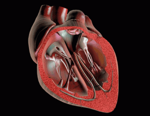
Cardiac Arrhythmia
 Cardiac arrhythmia, also called cardiac dysrhythmia or simply irregular heartbeat, is a modification of the normal heart beat. In other words, you have heart arrhythmia when your heart beat is too fast, too slow or irregular. The disease is not life threatening, but it can significantly impair the quality of your life if left untreated.
Your heart beats are assured by a natural pacemaker called sinus node (also kwon as sinoatrial node or sinuatrial node), a group of cells located in the upper wall of the right atrium of the heart, near the end of the superior vena cava. It works permanently as a regulator to control the heart rate. However, due to certain pathological circumstances, the sinus node can stop working normally, leading chaotic heartbeats. However, a simple and passage irregular heartbeat does not mean arrhythmia. The heart rate tends to vary depending on time of day, activity, emotional state, and taking of certain medications. You are diagnosed with cardiac dysrhythmia when the change becomes relevant, and leads to discomfort or symptoms.
Cardiac Arrhythmia may be of physiological origin or secondary to a disease. Whatever the causative factor, the disease is not so life threatening, unlike many other heart diseases. However, if left untreated, cardiac dysrhythmia can predispose you to various heart problems including stroke.
Cardiac Arrhythmia Symptoms
To better describe the symptoms of arrhythmia, it is important to enumerate the types of arrhythmia; there are two main types of arrhythmia:

Bradycardia – bradycardia is a medical term referring to a sudden and temporary decrease in heart rate; the heart beats less than 60 times per minute. Very often, it is characterized by slow pulse without any major symptoms. Sometimes, however, the disease causes a lack of blood flow to the brain and coronary arteries, and lead to:
- Fatigue
- Shortness of breath
- Dizziness.
Tachycardia – tachycardia is the opposite of a bradycardia; instead of slowness of heart beat, there is an abnormally fast heartbeat.
The normal heart rate varies normally around 60 beats per minute in an adult. Nevertheless, some physical factors such physical exercise, anxiety, fever, and consumption of alcohol may increase the number of your heart beats; that’s normal.
High acceleration of the heart rate without an accelerating factor will increase the oxygen requirements of the heart muscle. Persistence insufficiency of oxygen supply may cause myocardial ischemia, which can be responsible for heart failure, chest pain and more.
As bradycardia, tachycardia may be totally asymptomatic for a long period of time. As the disease worsens however, you can experience:
- Rapid heartbeat
- Shortness of breath
- Weakness
- Fainting
- Paleness, clammy skin
- Palpitations
- Chest pain, discomfort, tightness or pressure (angina pectoris).
Tachycardia may give rise to several medical conditions; vary depending on the group of cells (tissue) affected.
When the disease affects the atria (auricular tachycardia), tachycardia may be responsible for:
- Atrial fibrillation – irregular and rapid heart rhythm originating from the atria, the upper chambers of the heart
- Atrial flutter – the second most common of tachycardia after atrial fibrillation; it is characterized by an abnormal heart rhythm that occurs in the atria of the heart
- Supraventricular tachycardia (SVT) – also known as paroxysmal supraventricular tachycardia (PSVT), SVT consist of rapid heartbeat of over 100 beats per minute
- Wolff–Parkinson–White syndrome (WPW) – increased electrical pathway in the heart that leads to palpitations, fainting, and shortness of breath
When it affects the ventricles (ventricular tachycardia), tachycardia may be responsible for:
- Ventricular tachycardia (VT) – rapid heart rhythm originating in one of the ventricles of the heart
- Ventricular fibrillation – a life-threatening abnormal and rapid contraction of the ventricles in the heart
- Long QT syndrome – a congenital heart disease characterized by an abnormality in the heart’s electrical system.
Complications
Although arrhythmia is not a life threatening disease, it can considerably increase your risk of having a stroke, heart attack or sudden death. In addition, arrhythmia symptoms are similar to those of a heart attack or stroke, see your doctor immediately or call 911 if you experience any sign related to heart attack or stroke.
Cardiac Arrhythmia Causes
Heart arrhythmia occurs when the sinus node sends pulses disorderly. The disease can also occur when impulses originate in another location of the heart replacing those of the sinus node. Sometimes, arrhythmia comes from a blockage in the transmission of electrical impulse from the atria to the ventricles, which can be due to many factors:
- Stress
- Hyperthyroidism
- Genetics (congenital arrhythmia)
- Normal aging of the heart muscle
- Heavy consumption of tobacco, alcohol, coffee or other stimulants
- Certain respiratory diseases such as chronic bronchitis, emphysema and pulmonary embolism (obstruction of a lung artery by a blood clot or fat)
- Taking diuretics and/or antiarrhythmic agents (medication used to suppress fast heart beat)
- Any disorder causing a decreased blood flow (ischemia) such as arteriosclerosis (hardening of the walls of the arteries or/and heart) and atherosclerosis (fatty cholesterol deposits on the arteries).
Risk Factors
If you suffer from cardiovascular disease, you are at risk of developing some form of arrhythmia, because damage to the heart can prevent electrical impulses reach the ventricles. In general, you are prone to arrhythmia if you have:
- High blood pressure
- Valvular heart disease
- Heart failure
- Abnormal heart valves
- Coronary artery disease (CAD)
- Previous heart attack that causes death or damage to the heart tissue
- Malfunctioning of the nodal tissue (tissue containing the sinus node)
- Cardiomyopathy (weakening of the heart muscle or a change in
heart muscle structure).
Diagnosis
Heart arrhythmia diagnosis is often started by a physical exam. Your physician will use a stethoscope to listen to your heart beat, and eventually detect the arrhythmia. In addition, to facilitate the process, you can be asked questions about your medical history and things that can trigger symptoms of a heart arrhythmia.
However, to confirm the diagnosis, you will be recommended to undergo certain medical exams:
Electrocardiogram (ECG) – this radiographic examination consists of recording the electrical impulses of the heart. During the exam, the technician will place small electrodes on your chest and connect them to an electrocardiograph. During the procedure, your cardiologist will be able to determine the form and severity of the arrhythmia. Since the symptoms of arrhythmia do not usually occur in the hospital, you may be recommended to continue the exam at home by using portable ECG devices. Whenever possible, the ECG should be done while you’re having the symptoms.
Holter monitor – the Holter monitor is an ambulatory device allowing you to continuously monitoring your heart or ECG for up to 48 hours. During the examination, a housing registration will be given to you. The device will be connected by electrodes attached to your skin by an adhesive. The machine has a button which you can activate if you feel symptoms (palpitations, chest pain, etc) while recording. Increasingly, there is a marker placed on the recording, allowing your doctor to make the diagnosis.
Event monitor – If you have sporadic arrhythmia, you may be recommended to carry a cardiac event monitor to observe your abnormal heart rhythms. The device allows you to record your heart’s electrical rhythm while you are at home. The results will be reviewed by your health care provider during your next medical visit.
Tilt Test – also called tilt table test, a tilt test is a diagnostic procedure commonly used in arrhythmia diagnosis. During the procedure, you will be asked to take several positions. Your physician evaluates the reaction of your cardiovascular system during the movements. Your physician will recommend this exam if you experience dizziness, lightheadedness and fainting which can indicate a cardiac or vascular anomaly.
Cardiac ultrasound – this is a noninvasive imaging technique used to diagnose cardiovascular diseases. It helps your physician to determines the size of each atrium and detect any hypertrophy or blockage in the heart muscle. Cardiac ultrasound is often associated with a Doppler exam
Doppler Ultrasound – this is a special imaging technique allowing your cardiologist to explore the intravascular and intracardiac blood flow such as arteries and veins in your heart, abdomen, arms, legs and neck. It is based on a physical phenomenon of ultrasound called “Doppler Effect”.
Cardiac MRI or cardiovascular magnetic resonance imaging (CMR) – although rare, your cardiologist can recommend a cardiac MRI to evaluate the structure and function of your cardiovascular system.
Cardiac Arrhythmia Treatment
To increase your chance of recovery, even before starting the arrhythmia treatment, it is necessary to:
- Avoid everything that can lead to stress
- Stop smoking and all substances harmful to your heart health: alcohol, coffee, unsaturated fats, etc.
- Adopt a healthy balanced diet rich in fruits and cruciferous vegetables
- Lose weight if you are overweight or obese
- Doing physical exercises moderately and regularly.
Your doctor will use drugs, surgery, implantable devices or a combination of all of them to treat your condition.
Surgical treatment
Medications – The arrhythmia can be treated with various drugs including calcium channel blockers, beta-blockers and digoxin (Lanoxin). The medications aim to slow the rate of the ventricular contractions to help your heart rate return to normal.
Ablation Therapy – This is a nonsurgical technique performed in an electrophysiology (EP) laboratory. During the procedure, one or many electrode catheters are introduced into the veins of your arms or legs and advanced into your inner heart. A special machine is then used to provide energy to the heart muscle. Ablation Therapy is mostly performed to treat supraventricular arrhythmias
Synchronized electrical cardioversion – this is a non surgical process by which an abnormally fast heart rate or cardiac arrhythmia is corrected. During the procedure, your cardiac surgeon delivers a therapeutic dose of electric current to the heart muscle at a specific moment. This method is used in the treatment of arrhythmia when the tachycardia originate from the top half of your heart (atria), including atrial fibrillation.
Vagal maneuvers – your doctor may recommend you to use vagal maneuvers to slow fast heart rate related to arrhythmia. The techniques stimulate the vagus nerve to result in slowed conduction of electrical impulses through the atrioventricular (AV) node of the heart. Some of common vagal maneuvers techniques include:
- Gagging
- Coughing
- Putting pressure on your eyelids
- Holding your breath and bearing down (Valsalva maneuver)
- Immersing your face in ice-cold water (diving reflex).
Implantable Devices
Pacemakers – in case of slow heartbeat, your surgeon may implant a pacemaker (battery). This device, once implanted in your body, delivers electrical pulses to the heart, which accelerates its functions when it is too slow. In general, the pacemaker implantation is performed under local anesthetic. You should take precautions when you wear a pacemaker; talk to your doctor for more information.
Implantable cardioverter defibrillator (ICDs) – a defibrillator is a type of pacemaker allowing the detection and treatment of tachycardia or ventricular arrhythmias. It is used as a preventive measure against sudden death, mainly in heart disease with mechanical malfunction of the left ventricle. However, ICD is not popular because the cost of this therapy is too high.
Surgical Treatment
Sometimes, all other therapeutic methods fail to produce satisfying results in the treatment of arrhythmia; surgery becomes the only option to consider. The two most common surgical procedures that your surgeon can perform are ventricular aneurysm surgery and coronary bypass surgery.
Ventricular aneurysm surgery – An aneurysm is a section of a defective wall that bulges outward. A ventricular aneurysm is a defect in the ventricle of the heart, usually produced by transmural infarction. If your surgeon suspects the arrhythmia is resulted from a ventricular aneurysm, he may remove the ventricular aneurysm surgically.
Coronary Artery Bypass Surgery– obstruction of a coronary artery reduces blood flow reaching the heart muscle. When blood flow is reduced, it can result in angina pectoris or even heart attack. Coronary Artery Bypass Surgery consist of installing a graft between the aorta (or an extracardiac artery) and the coronary artery to restore normal blood flow in a narrowed or obstructed artery.
Cardiac Arrhythmia Prevention
To prevent all forms of arrhythmias or their complications (if you already have the disease), practice the following preventive methods:
- Do not smoke or quit smoking
- Eat a heart-healthy diet rich in omega-3
- Avoid caffeine, alcohol and sugar (in all its forms)
- Exercise regularly – fast walking is excellent
- Avoid stress, depression and anxiety
- Lose weight if you are obese or overweight
- Take your medications as prescribed if you have a cardiovascular related problem.
References:
1 – Heart arrhythmias; Treatments and drugs: mayoclinic.com
2 – Catheter ablation therapy; McGraw-Hill Concise Dictionary of Modern Medicine: The McGraw-Hill Companies, Inc.
3 – Vagal maneuvers for a fast heart rate: Robin Parks, Caroline S. Rhoads, MD – Internal Medicine; Stephen Fort, MD, MRCP, FRCPC – Interventional Cardiology
4 – Ventricular Aneurysms: Vibhuti N Singh, MD, MPH, FACC, FSCAI, Director, Suncoast Cardiovascular Center; Chair, Cardiology Division and Cath Labs, Department of Medicine, Bayfront Medical Center; Clinical Assistant Professor, Division of Cardiology, University of South Florida College of Medicine
5 – https://health.google.com/health/ref/Cardiomyopathy
6 – “What Is Arrhythmia?”. http://www.nhlbi.nih.gov. July 1, 2011. Retrieved 7 March 2015.
7 – “What Are the Signs and Symptoms of an Arrhythmia?”. http://www.nhlbi.nih.gov. July 1, 2011. Retrieved 7 March 2015.
8 – “Types of Arrhythmia”. http://www.nhlbi.nih.gov. July 1, 2011. Retrieved 7 March 2015.
9 – Martin, C; Matthews, G; Huang, CL (2012). “Sudden cardiac death and Inherited channelopathy: the basic electrophysiology of the myocyte and myocardium in ion channel disease.”. Heart 98: 536–543. doi:10.1136/heartjnl-2011-300953. PMID 22422742.
10 “How Are Arrhythmias Diagnosed?”. http://www.nhlbi.nih.gov/. July 1, 2011. Retrieved 29 March 2015.
11 – “How Are Arrhythmias Treated?”. http://www.nhlbi.nih.gov/. July 1, 2011. Retrieved 10 January 2015.
12 – “Who Is at Risk for an Arrhythmia?”. http://www.nhlbi.nih.gov/. July 1, 2011. Retrieved 1 May 2015.