Table of Contents
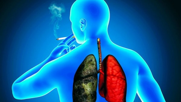
Human being has two lungs, one at left and the other one at right. They are located on the diaphragm and protected by the rib cage. The left lung is divided into two lobes; the right lung has three lobes, which makes it larger and heavier. All these lung tissues are formed by a group of cells that allow the exchange of vital gases, oxygen and carbon dioxide. At every moment, thousands of cells naturally die to be replaced by new healthy cells. Lung cancer occurs when a group of cells refuses to die and begin to multiply uncontrollably to form malignant growth in the tissue of the lungs.
 Smoking is the leading cause of small cell lung cancer. It is responsible for about 90% of lung cancers among men and 78% among women. It is estimated that nearly 98% of small cell lung cancer patients have a smoking history. In addition to direct use of tobacco, being frequently exposed to secondhand smoke or carcinogenic particles in the air – asbestos, radon gas, gasoline, etc. – can also cause small cell lung cancer.
Once inhaled, toxins contained in tobacco smoke (including secondhand smoke or passive smoke) accumulate in the lungs and begin their degenerative effects, slowly and sometimes asymptomatically. When symptoms finally emerge, the tumor is already very advanced. Approximately 65-70% of patients with small cell lung cancer have an extensive malignant tumor at diagnosis. In general, extensive-stage small-cell lung cancers are incurable; they kill their victims within 12 months after diagnosis.
Small Cell Lung Cancer Risk Factors
Most common small-cell lung cancer risk factors include:
- Being a miner – mining uranium frequently increases the risk of all types of lung cancers including small-cell lung cancer. The risk is higher if you are a smoker.
- Radiation – exposure to tobacco smoke and radiation are risk factors that act synergistically to the development of small-cell lung cancer.
- Chloromethyl ether – studies has shown thatbis(chloromethyl) ether can cause lung cancer and other malignant tumors in people and animals. The Department of Health and Human Services (DHHS) has determined that bis (chloromethyl) ether is a known human carcinogen.
- Carcinogenic substances – prolonged exposure to certain substances such as radon and asbestosis can lead to the development of small-cell lung cancer. Asbestos exposure alone increases your lung cancer risk by 9 times; when associated with cigarette smoking, the risk can be increased up to 50 times.
- Diet low in fruits and vegetables – diet low in fruits and vegetable can lead to major health problems. In the other hand, regular consumption of fruits and vegetables exert a protective effect in people exposed to tobacco smoke. Numerous studies have shown a lower risk of lung cancer among consumers of fruits and vegetables rich in beta-carotene: sweet potatoes, pumpkins, carrots, spinach and other dark green vegetables, winter squash, etc.
- Sex – men are twice affected by lung cancer than women are; however, according to the National Cancer Institute, the incidence of lung cancer started to decline among males in the early 1980s and has continued to do so over past 20 years.
- Age – lung cancer can affect people of all ages; however, the most cases of small cell lung cancer occur in people aged 35-75 years. According to American Cancer Society, the incidence of lung cancer (non-small cell lung and small cell combined) among men and women are as follows:
| Age | Men | Women |
| 0-39 | 0.04% | 0.03% |
| 40-59 | 1.24% | 0.92% |
| 60-79 | 6.29% | 4.04% |
| from birth to death | 8.09% | 5.78% |
Small Cell Lung Cancer Symptoms
Due to its aggressive characteristic, small cell lung cancer is rarely asymptomatic; it is often associated with a variety of symptoms. Small cell lung cancer tends to cause cough, fatigue and anorexia. With times, however, other symptoms can emerge, which vary depending on tissue affected.
Symptoms due to primary tumor
- loss of appetite
- weight loss
- chest pain
- shortness of breath
- Coughing up of blood or of blood-stained sputum (hemoptysis).
When the cancer has reached surrounding organs/tissues, it can provoke:
- wheezing, due to compression of the tracheaand mainstem bronchi;
- superior vena cava obstruction
- harsh, raspy, or strained voice (hoarseness)
- phrenic nerve palsy
- Difficulty swallowing (dysphagia), due to compression of esophagus.
Symptoms associated with distant metastases can include:
- neurological dysfunction, due to brain metastasis, spinal cord compression
- bone pain, due to bone metastasis
- Abdominal/right upper quadrant pain, due to liver metastasis.
Small Cell Lung Cancer Complications
Small cell lung cancer is often subject to complications. Even after treatment, the tumor can obstruct the airways and increases the risk of respiratory infections like bronchitis or pneumonia.
In addition, the cancer can spread into other parts of the body to form metastases. Most often, the metastases are formed in the bone, brain or liver. Once in these organs, the tumor can cause bone disease, neurological problems, liver disease, and more serious health problem including death.
Non-Small Cell Lung Cancer Diagnosis
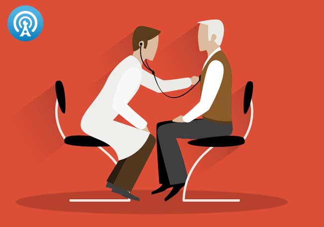
To begin the diagnosis, your doctor will do a physical examination of your body to search for signs indicating non-small cell lung cancer. He can use a stethoscope to listen to the sound of your breathing to determine how your lungs function. He can also ask you to inhale very deeply, and tap on your chest. In addition, you will be asked about your medical history and the characteristic of the symptoms you experience.
However, to confirm a non-small cell lung cancer, several tests must be performed. In general, your oncologist can recommend complete blood count (CBC), spectrum test, imaging techniques, liver function tests and biopsy.
Sputum Test – a sputum culture can be the first test recommended by your physician. It is the easiest way to detect and identify bacteria or fungi that infect the lungs or the airways. However, although useful, sputum test cannot confirm you have non-small cell lung cancer; other diagnostic procedures must be performed
Complete blood count (CBC) – this exam aims to analyze your red cells, white cells and platelets. It is a very simple procedure during which a nurse takes sample of your blood for laboratory analysis. Usually, the sample obtained is analyzed by a lab specialist who measures the number of red blood cells, hemoglobin and hematocrit, the volume of cells circulating in the blood compared to the total volume of blood. In addition, the CBC used to calculate the MCV (mean corpuscular volume), the MCHC (corpuscular hemoglobin concentration) and the MCH (Mean corpuscular hemoglobin).
Bone scan – this imaging technique allows your doctor to detect very early pathogenic changes in your bones, sometimes not even visible on standard x-rays. During the procedure, the specialist will inject a small amount of radioactive substance which will bind to the diseased bone, and gives off radiation. The radiation emitted is detected by a gamma camera that create picture of your bones. The purpose of this test is to determine if the cancer has spread to any bone in your body.
Ct scan – a scanner is the use of x-rays to create images of your internal organs. It can detect abnormalities not visible on standard x-rays and ultrasound. The CT scan allows not only to detect the primary cancer in your lungs but also to highlight lymph nodes or/and liver metastases.
Magnetic resonance imaging (MRI) – with this imaging technique, your health care provider can visualize organs inside of your body, and detect disease. In the diagnosis of small cell lung cancer, your doctor can visualize and analyze the structure of your lungs, to look for abnormalities, inflammation, and presence of a tumor. The MRI not only allows your physician to detect the cancer but to know the exact size and extent of the tumor.
Bronchoscopy – this is a medical procedure used to examine the interior of the airways. During the test, your physician introduces a thin and flexible camera (bronchoscope) into the air passages of your lung to search benign or malignant conditions. A bronchoscopy can be performed for therapeutic or diagnostic purposes.
Chest x-ray – a chest x-ray is a painless diagnostic procedure that about 10 minutes. It creates pictures of your thoracic cage, which allow your health care provider to detect abnormalities in your lungs, trachea, bronchi and layers surrounding the lungs (pleura). This procedure cannot give specific details on the cancer, but it can reveal abnormal tissue growth.
Ultrasound – during this imaging technique a medical technician uses painless high-frequency sound waves to visualize different organs of your body including your lungs. It involves applying an ultrasound sensor (transducer) on your chest in order to obtain images of your lungs. Images obtained will be sent to your doctor or an ultrasound specialist who will declare whether or not you have a tumor in your lung.
Liver function tests – this is a group of tests that are used to evaluate the function of the liver. Usually, a medical technologist will perform those tests to determine if the cancer has spread to your liver.
Positron emission tomography (PET) scan – this imaging technique gives your doctor an idea on how your tissues and organs are functioning. During the test, a radioactive tracer is injected into your body which will accumulate on the diseased tissue. Usually, the location where the tumor is located shows up as brighter spots on the PET scan. A PET differs from conventional X-rays and MRI; it can detect the tumor at an earlier stage.
Thoracentesis – also known as pleural TAP, thoracentesis is an invasive procedure involves draining fluid or air from your pleural cavity. Thoracentesis may be performed for diagnostic purposes, removing fluid for examination; or a palliative treatment – removing fluid to improve lung function. During the procedure, a cannula or hollow needle is carefully injected into your chest to remove the liquid; usually after administering a local anesthesia.
Biopsy – to accurately confirm a non-small cell lung cancer diagnosis, your doctor will takes sample from the tumor to microscopically examine it. This microscopic study is done to obtain accurate information on the overall structure of the fragment removed. The biopsy is important to confirm with certainty the presence of cancer cells in your lungs. In general, your physician will perform CT scan-directed needle biopsy, mediastinoscopy with biopsy, open lung biopsy or pleural biopsy.
Lung Cancer Stages
Once the cancer is found in your lung, it is important for your doctor to determine its stage. The staging is necessary in the choice of the treatment and evaluation of the prognosis. In general, lung cancer includes the following stages:
Stages of non-small cell lung cancer
- Stage I – a stage 1 lung cancer is much localized in the lung; the tumor has affected the underlying lung tissue, but has not spread into nearby lymph nodes. The survival chance is high.
- Stage II – cancer has affected the underlying lung tissue and has spread into lymph nodes surrounding the lungs.
- Stage IIIA – the cancer has spread into other lymph nodes or tissues surrounding its initial location in the chest cavity.
- Stage IIIB – at this stage, le cancer has invaded not only the chest cavity, but also other vital organs: heart, blood vessels, trachea and/or esophagus.
- Stage IV – Stage IV indicates a very serious phase of the tumor. The cancer, from lungs and surrounding organs, has spread into other organs such as the liver, bones or brain; survival chance is very low.
Stages of small cell lung cancer
- Limited – the cancer remains in the thorax and has affected one lung;
- Extensive – the tumor has spread to other organs outside the thorax, and most often, both lungs are affected.
Non-Small Cell Lung Cancer Treatment
Most lung cancers including non-small cell lung cancer are incurable; the treatment aims at shrinking the tumor to prevent complications and relieve the symptoms in order to help patients live better and longer. Along with a healthy lifestyle, the therapies can help you live for years without major complications.
However, after treatment, non-small cell lung cancer can recur or relapse any time; therefore, even if you feel good during the remission, it is important that you see your doctor regularly to evaluate your health.
To determine an appropriate treatment, your oncologist will consider your general health, age, and most importantly the stage of the tumor. In general, non-small cell lung cancer is treated with one or an association of the following therapies:
Surgical treatment
To have complete access to your lungs, your surgeon may perform a thoracostomy, a major surgical intervention performed under general anesthesia. Your surgeon will open your chest wall or does incision between your ribs to fully expose your lungs. During the surgery, he removes part or the entire diseased lung. Depending on the extension of the tumor, the surgeon can also remove nearby lymph nodes. The goal of the surgery is to remove as much cancerous tissue as possible to reduce symptoms and help you live longer.
Surgery is the preferred treatment of non-small cell lung cancer stages I and II. Patients who have stage IIIB or IV cancer associated with pleural or neoplastic effusion are not candidates for surgery. The surgery should be performed in the absence of contraindications such as evidence of spread of the tumor outside the lungs, endobronchial tumor located too close to the trachea, and other serious illnesses: coronary artery disease, or respiratory failure due to chronic obstructive pulmonary disease (COPD).
Lung Cancer Chemotherapy
Chemotherapy is a cancer treatment consists of using strong chemical agents to destroy cancer cells or prevent them from multiplying. Chemotherapy drugs can be administered orally or intravenously. Similarly, chemotherapy treatment may consist of a single chemotherapeutic agent (monochemotherapy) or several chemotherapeutic agents (polychemotherapy).
The use of chemotherapy to treat a stage I or II non-small cell lung cancer can sometimes bring good results. When the chemotherapy is administered preoperatively and before radiotherapy, it can significantly reduce the tumor mass and increase remission and overall survival. Used after surgery, chemotherapy drugs attack and destroy cancer cells remaining from the surgery.
Radiation Therapy (Radiotherapy)
Radiotherapy involves exposing cancer cells to ionizing radiation that alter the composition of their genetic information. Unlike chemotherapy, radiation acts locally on the region that is irradiated, thereby limiting its action to the tumor and a small surrounding tissue. Radiotherapy may be used before or after surgery, alone or in combination with chemotherapy.
In the treatment of non-small cell lung cancer, radiotherapy is sometimes used instead of surgery when the thoracotomy is contraindicated due to cardiopulmonary failure or other serious illness. Radiation therapy can bring good results in reducing bone pain associated with the tumor. In addition, radiotherapy can be very useful in some types of tumors resulting in:
- superiorvena cavaobstruction (SVCO)
- spinal cord compression
- brain metastases
- spitting of blood (hemoptysis)
- Bronchial obstruction.
Laser therapy
If you have a non-metastatic non-small cell lung cancer, your oncologist can use high-intensity light to shrink or destroy the tumor. This therapy cause less adverse effects, but it can only be used to treat superficial cancers.
Other therapies
In addition, bronchodilators, oxygen therapy, and physiotherapy may be necessary in cases of bronchial obstruction. Antibiotic therapy can be recommended in case of superinfection (an infection following a previous infection).
Non-Small Cell Lung Cancer Survival Rates
Non-small cell lung cancer prognosis is often alarming; the cancer is often diagnosed at an advanced stage, which makes treatment very often ineffective. The overall 5-year survival rate is approximately 13% – 15%. Without treatment, victims of non-small cell lung cancer do not survive one year. Patients with a stage 1 cancer well confined to the lung, five-year survival rate is more or less better, about 70%. Patients with stage III non-small cell lung cancer five year survival rate is about 15%.
However, the survival rates tend to differ from one race to another. According to the National Cancer Institute, for 1999-2005, five-year relative survival rates by race and sex were:
- 13.7% for white men;
- 18.3% for white women;
- 10.8% for black men;
- 14.5% for black women.
Non-Small Cell Lung Cancer Prevention
Stop Smoking –  -Although there are treatments for lung cancer, its prevention is the ideal option. Smoking causes almost 90% of lung cancers; quitting smoking remains the safest lung cancer prevention. Whether you are a victim of lung cancer or want to prevent it, from now, you need to:
- stop smoking (including secondhand smoke)
- eat a healthy diet rich in fruits and cruciferous vegetables
- exercise regularly
- sleep well
- Clean the air your breath.
Stopping smoking is not easy, but you will succeed with determination and discipline. You can use certain products such as nicotine patches, quit smoking pills or tablets, and stop smoking supplements. In addition, you can get support from parents, friends, and social groups. Doing the right thing is never easy, but with determination you can; make a decision to stop smoking today.
Early detection – thanks to an early screening technique called DNA methylation profiling, researchers are now able to identify molecular indices of lung cancer in their genesis. Methylation is an epigenetic process, causing diseases and reversible changes of gene expression. These abnormalities, if left untreated, can lead to the development of lung cancer.
Avoid exposure to carcinogens – certain substances such as asbestos or other air pollutants can cause genetic mutations leading to lung cancer. As long as it is possible, avoid any work that exposes you to asbestos which mostly found in mines and some old buildings. In addition, avoid prolonged exposure to radon (estimated to cause about 21000 lung cancer deaths per year, according to EPA), chromium hexavalent (CrVI) compounds, propane etc. The following substances, although the risk is minimal, can also cause lung cancer if you inhale them very often:
- arsenic
- beryllium
- vinyl chloride
- nickel chromate
- mustard gas
- diesel exhaust
- Talcum powder.
Avoid air pollution – many studies have shown that some pathogenic pollutants suspended in the air are responsible for nearly 5% of deaths from cancer of the trachea, bronchi and lungs. These particles can be originated from combustion of coal, oil, natural gas, incineration of waste materials, and much more. Therefore cleaning the air you breathe can also help your lungs to remain healthy.
Fruits and cruciferous vegetables – consuming a diet rich in fruits and cruciferous vegetables have preventive effects on all types of cancer. It is shown that people who eat a daily variety of fruits and vegetables have fewer problems related to free radicals. It is also shown that there is a lower risk of cancer among consumers of fruits and vegetables rich in beta carotene such as sweet potatoes, broccoli, pumpkin, carrots, spinach, winter squash, etc.). Even smokers – including second hand smokers- can beneficiate from regular eating of fruits and cruciferous vegetables.


