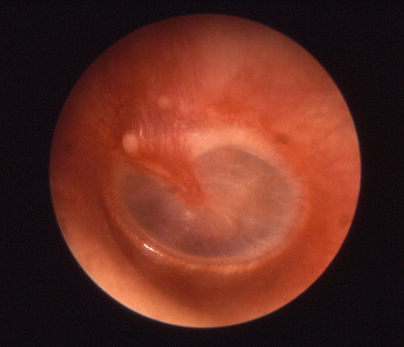
Ear is the human organ of hearing and balance; It collects and analyzes sounds. When a noise is made, the ear converts those sound signals into electrochemical impulses, and then sends them to the brain. This physiochemical process is accomplished by three main parts of the ear: Outer ear, Middle ear, and Inner ear
The outer ear, also called pinna, is made of the auditory canal and the eardrum. The role of the outer ear is to collect sounds and provide protection for the middle ear in order to prevent damage to the eardrum. The auditory canal is lining by a very thin mucous membrane (mucosa). This mucous produces a natural substance called earwax. The major function of the earwax is to support the skin inside the ear canal and prevent dirt and other pathogenic substances from penetrating the middle and inner ear.
The middle ear is an air-filled cavity containing the eardrum, cubical cavity (separated from the external ear by two small membranes), the round window and the oval window (both approximately 2.5 mm2 in size). Between the eardrum and the oval window are located three auditory ossicles called successively hammer, anvil, and the stirrup. In addition, the middle ear also contains small cavities in the mastoid (bone behind the ear) and the Eustachian tube, canal linking the eardrum to the nasopharynx (part of the pharynx behind the nasal passages).
The inner ear includes the cochlea (the organ of hearing) and the labyrinth, in which found the membranous labyrinth. These two organs contain two fluids, the endolymph (within the labyrinth) and the perilymph (within the cochlea). The liquids are attuned to the effects of gravity and motion, and playing a major role in regulating electrochemical impulses of hair cells.
Unfortunately, the functions of the ear (outer ear, middle ear, and inner ear) can be impaired with either infections or injuries. One of the most common ear problems is otitis.
Otitis is infection or inflammation of the ear cavities. There are two common types of ear infections, otitis media and otitis externa.
Ear Infection Causes and Risk Factors
Otitis media can be acute, secretory or chronic depending of its evolution and causes.
Acute otitis media is an inflammation caused by viral or bacterial infection, Streptococcus pneumoniae, Haemophilus influenza. It affects mostly children from six months to two years, and particularly children who grow up in community. The infection is first pharyngeal, and then spreads to the inner ear through the Eustachian tube.
Secretory otitis media – also called otitis media with effusion (OME), is usually an inflammation of the middle ear accompanied with a fluid effusion (without pus) due to malfunction of the Eustachian tube. It is characterized by a repeated acute otitis and/or a decrease of hearing. Secretory otitis media is a leading cause of hearing loss in children.
Chronic otitis – Chronic otitis media is the Inflammation of the skin lining the middle ear canal due to repeated blockage of the Eustachian tube. This blockage can be caused by allergies, repeated infections, ear trauma, untreated acute otitis or eardrum perforation. Chronic otitis results in discharge, decrease of hearing, and sometimes hearing loss. Complications can lead to facial paralysis, meningitis, cholesteatoma (development in the inner ear of a benign growth called ear cyst), mastoiditis (infection or inflammation of the adenoids) or epidural abscess (life-threatening inflammation around the brain).
Otitis externa – otitis externa is a common type of infection of the external ear canal caused by viral or bacterial infection. It has less chance of complications.
Ear Infection Symptoms
The main symptoms of otitis media include ear pain, itching, and discharge. The pain can severe and associated with fever of about 38.5 ° C, earache, tinnitus (ringing or buzzing in the ear) and sometimes vomiting.
Among infants, symptoms may include irritability, crying, tugging or rubbing of the ear, digestive disorders and loss of appetite.
In the absence of treatment, otitis can lead to: congestive otitis, the eardrum becomes red; otitis catarrhal, the eardrum becomes smooth and opaque; Purulent otitis, bulging eardrum with presence of pus; perforated ear infection, rupture in the eardrum causing temporary hearing loss and occasional discharge of pus.
Diagnosis of otitis
Mild Acute otitis can be diagnosed by physical exam and evaluation of the clinical signs of the ear, usually redness and inflammation. If there is discharge of pus, visual observation can be complemented with tympanometry, exam used to test and measure the pressure on both sides of the tympanic membrane); auriscopy (examination of the ear by the aid of the auriscope to detect abnormalities) and CT scan of the head or mastoids, which allows your physician to detect presence or the effects of the infection in the mastoids.
Ear Infection Treatment
The treatment of acute otitis is antibiotherapy. If there is fluid behind your eardrum, antibiotics can be associated with paracentesis, removal of fluid from the eardrum cavity with a needle. The treatment of Secretory otitis is, depending on complications, antibiotherapy, surgical removal of the adenoids (Adenoidectomy) or installation of an aerator transtympanic. For externa otitis, most doctors recommaend antiseptic, antibiotic or antifungal drops.
Alternative treatment
Drink lots of carrot and lemon juice, Eat fresh watercress
Drops into each ear a few drops of warm onion juice
Put salt in a sock/cloth, heat it (not too hot please), then Apply on the infected ear
Table
| Causes | Symptoms And Signs | Prevention | Treatment | ||||
Tobacco smoke or other irritants |
|
|
Conventional Treatment
Natural Alternative · Drops into each ear a few drops of warm onion juice
|
||||
References:
http://en.wikipedia.org/wiki/Ear#Parts_of_the_ear
Appropriate Use of Antibiotics for URIs in Children: Part I. Otitis Media and Acute Sinusitis by SF Dowell, M.D., M.P.H., B Schwartz, M.D., WR Phillips, M.D., M.P.H., and The Pediatric URI Consensus Team (American Family Physician October 1, 1998, http://www.aafp.org/afp/981001ap/dowell.html


